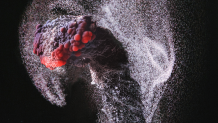
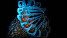
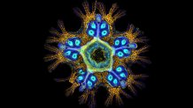
An eldritch abomination – or a baby starfish magnified twenty times its size?
Photos from this year's Nikon's Small World Photomicrography competition show a surreal, almost dizzying look at the natural world from underneath the lens of a light microscope. Cells taken from the human body transforms itself into a Lovecraftian landscape in this contest, while slime – disgusting in real life – turns into a work of art under the hands of a researcher.
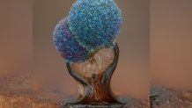
This year's competition is Nikon's 48th celebrating the art and technical science of photomicrography. The first place winner, an image of a three millimeter long hand of a gecko embryo, was stitched from hundreds of images in order to capture the details of nerves, cells and bones.
See the full list of winners and honorable mentions here.
Grigorii Timin & Dr. Michel Milinkovitch / Nikon Small World
First place winner: the hand of a Madagascar giant day gecko embryo, taken at 63x magnification.
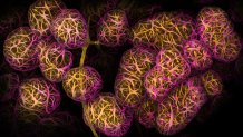
Dr. Caleb Dawson / Nikon Small World
Second place winner: breast tissue showing cells responsible for human milk production (alveoli) at 40x magnification.
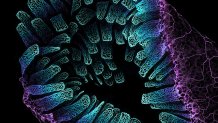
Satu Paavonsalo & Dr. Sinem Karaman / Nikon Small World
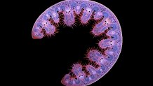
Third place winner: an adult mouse's intestine, showing its blood networks at 10x magnification.
Alison Pollack / Nikon Small World
Slime mold shown at 10x magnification.
Ole Bielfeldt / Nikon Small World
The extinguished wick of a candle showing particles of carbon being released at 2.5x magnification.
Murat Öztürk / Nikon Small World
A tiger beetle clamps down on a fly at 3.7x magnification.
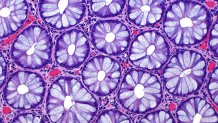
Dr. Ziad El-Zaatari / Nikon Small World
Crypt cells from a human colon at 20x magnification.
Anatoly Mikhaltsov / Nikon Small World
A cross section of a dune grass leaf, taken at 10x magnification.
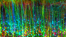
Dr. Andrea Tedeschi / Nikon Small World
The motor area of the brain of a genetically modified mouse after suffering a brain injury.
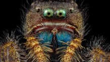
Dr. Andrew Posselt / Nikon Small World
A bold jumping spider at four times the magnification.
Dr. Andrew Moore / Nikon Small World
Human cells in different stages of mitosis at 100x magnification. Chromosomes are in orange.
Dr. Eugenijus Kavaliauskas / Nikon Small World
The face of an ant, shown at five times the magnification.
Frank Fox / Nikon Small World
A hibiscus flower with pollen spores, taken at 10 times the magnification.
Dr. John Hart / Nikon Small World
Amino acid crystals, seen at 20 times the magnification.
Ye Fei Zhang / Nikon Small World
A butterfly egg, seen at 10x magnification.
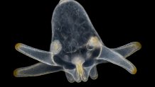
Wim van Egmond / Nikon Small World
The larva of a sea anemone, at 6.3 times the magnification.
Michael Landgrebe / Nikon Small World
A moss spore capsule, seen at 20x magnification.
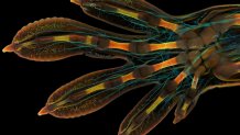
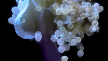
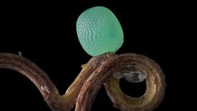
Dr. Laurent Formery / Nikon Small World
A two-month old starfish, seen at 20x magnification.
Karl Gaff / Nikon Small World
A freshwater midge larva at 10 times the magnification.
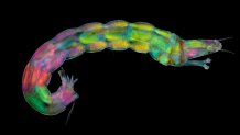
Danny J. Sanchez / Nikon Small World
Etch tube in Brazilian quartz at six times the magnification.